Medical imaging is one of the most critical tools in modern healthcare. From X-rays and CT scans to MRIs, ultrasounds, mammograms, and pathology slides, these images carry essential information that directly shapes how a patient is diagnosed and treated.
Detecting disease at an early stage often depends on how quickly and accurately a scan is reviewed. As imaging volumes grow across hospitals and diagnostic centers, so does the pressure on clinical teams to deliver consistent, timely results.
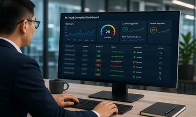
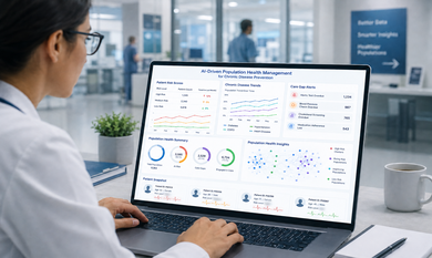
AI-powered medical imaging analysis is changing how diagnostic workflows operate. Not by replacing doctors or radiologists, but by helping them process scans more efficiently, surface patterns that need attention, and prioritize cases that require faster review.
AI acts as a support system for medical professionals, giving them better tools to work more consistently and at greater scale. The final diagnosis always remains the responsibility of qualified healthcare experts.
The Real Challenge: More Scans, More Complexity, and Less Time
Diagnostic imaging demand has grown significantly. Preventive screenings, chronic disease management, emergency care, and population health programs all depend heavily on imaging. Radiologists and diagnostic teams are expected to review more scans, faster, while maintaining high accuracy.
The manual review process has real limits. A radiologist reading hundreds of scans in a single shift faces mental fatigue, especially when looking for subtle, early-stage findings. Low-contrast lesions, small nodules, or faint tissue changes are more likely to be missed under time pressure and heavy workloads.
This is where medical imaging AI solutions can provide meaningful clinical support. Instead of slowing diagnostic teams down, well-designed AI can automate repetitive analysis steps, organize imaging queues by priority, and highlight scans with suspicious regions for faster review.
Early-stage disease detection is most effective when imaging workflows are structured, consistent, and supported by tools that help clinicians focus their expertise where it matters most.
How AI Analyzes Medical Images
AI does not view medical images the way a human does. It processes them computationally, using layers of trained models to identify patterns within pixel and voxel data.
The foundation of AI in diagnostic imaging is deep learning, where models are trained on large sets of labelled medical scans. Over time, a model learns to associate specific visual features with specific clinical conditions, such as the shape of a nodule, the texture of tissue, or irregular boundaries within an organ.
Computer vision techniques allow the system to perform tasks like image segmentation (separating different structures within a scan), object detection (locating a region of clinical interest), anomaly scoring, and risk stratification. Pattern recognition enables AI to compare new scans against thousands of previously labelled examples.
Healthcare AI software built for radiology can support scan prioritization, side-by-side comparison of current and historical scans, image classification, and flagging of regions that may need clinical review. These outputs are designed to inform and assist, not replace, a radiologist's clinical judgment.
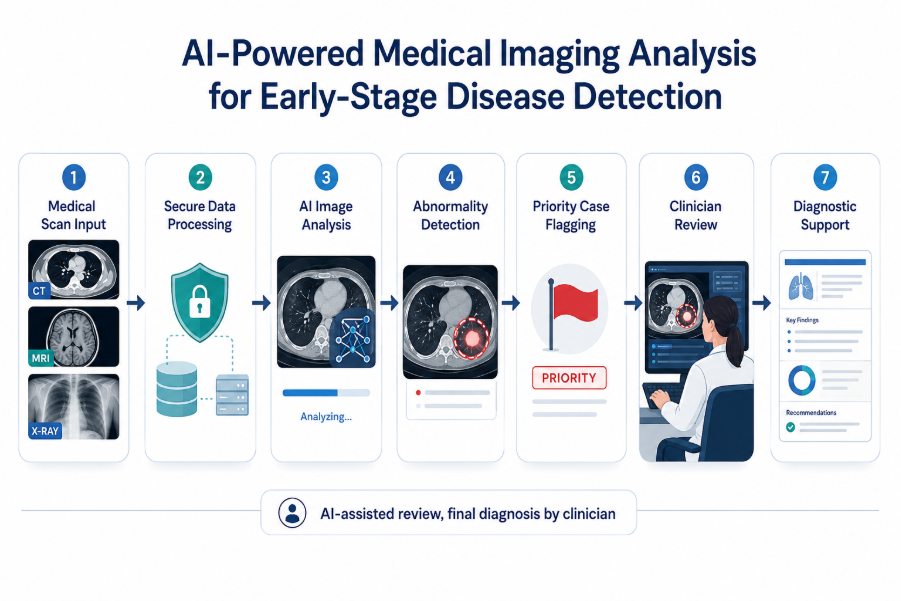
Figure: AI-powered medical imaging workflow for scan analysis, abnormality detection, priority flagging, and clinician review.
Practical Use Cases of AI in Medical Imaging
AI imaging tools are being explored across several clinical specialties. Below are some of the most active areas of application.
Cancer Screening and Tumor Detection
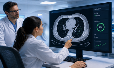
In mammography and lung CT imaging, AI can assist by highlighting potential masses, calcifications, or nodules for radiologist review. In tumor monitoring workflows, AI can compare scan sequences over time and flag changes in size or density that may warrant closer clinical attention.
For example, consider a high-volume diagnostic center where an asymptomatic patient undergoes a routine chest CT. A very small pulmonary nodule may be difficult to notice during a busy review queue, especially when scan volumes are high. An AI-assisted imaging system can flag the suspicious region, move the case higher in the review queue, and help the radiologist examine it with more attention. The final decision still remains with the clinician, but this type of support can help healthcare teams act earlier when follow-up is needed.
Neurology and Brain Imaging
AI models trained on brain MRI and CT data can support the identification of hemorrhages, lesions, or structural irregularities. In time-sensitive situations such as suspected stroke, AI can help prioritize scan queues and surface findings that need faster clinical review.
Cardiology Imaging
Cardiac imaging analysis with AI can support the review of echocardiograms and cardiac CT scans. AI tools can assist in identifying structural irregularities, measuring cardiac parameters, and organizing imaging reports for cardiologist review.
Orthopedic and Bone Imaging
AI tools trained on musculoskeletal imaging can assist in detecting fractures, joint degeneration, and alignment concerns in X-rays and MRIs. This helps orthopedic teams reduce manual review time in high-volume settings.
Chest and Respiratory Imaging
Chest X-ray analysis remains one of the most widely developed applications of AI in radiology. AI can assist in identifying opacities, consolidations, and respiratory abnormalities, helping triage imaging queues where scan volumes are high.
Healthcare organizations exploring AI healthcare solutions in Canada can use medical imaging intelligence to support faster review, better prioritization, and more structured diagnostic workflows.
What Happens Behind an AI Imaging System
Building a medical imaging AI system involves several interconnected technical and clinical layers.
It starts with secure image data collection, followed by data cleaning and standardization to ensure consistency across different imaging equipment and formats. Medical image annotation by trained clinical experts is a critical step, since labelled data is the foundation for model accuracy.
Computer vision in healthcare involves training deep learning models on annotated image datasets, then validating those models against unseen clinical data before any patient-facing deployment. Model performance is measured across different patient groups, imaging conditions, and equipment types.
On the integration side, a well-built system connects with PACS (picture archiving and communication systems), EHR and EMR platforms, and diagnostic reporting tools. Clinicians interact with AI medical image analysis outputs through structured dashboards that display heatmaps, bounding boxes, or segmentation overlays, which visually indicate regions the model has flagged for review.
Audit logs and traceability are built into the system to ensure every AI output is recorded. Security layers protect sensitive patient imaging data in line with applicable healthcare privacy regulations. These components together ensure the system is technically functional and clinically responsible.
Why Human Review and Clinical Validation Are Essential
AI is a tool that supports clinical decision-making, not one that replaces it. This distinction is both ethically and practically critical.
Even well-trained AI models can produce false positives or miss findings in unusual or underrepresented cases. Clinical validation is the process of testing a model's performance in real-world conditions before and after deployment. Validation should cover different imaging equipment, diverse patient populations, and a range of clinical presentations.
Explainability matters as well. Clinicians need to understand why the AI flagged a region, not just that it did. Visual tools like heatmaps and overlay markers help doctors review AI outputs with context and make informed judgments.
Model monitoring after deployment ensures performance does not degrade over time. Changes in imaging equipment, patient demographics, or clinical protocols can affect how an AI model behaves in practice.
Responsible AI adoption in healthcare means building systems where final clinical judgment remains with qualified medical professionals, and where AI serves as a structured, transparent, and auditable support layer.
For healthcare organizations in Canada, AI imaging systems should also be planned with privacy, security, clinical validation, and applicable healthcare technology requirements in mind. This includes careful handling of patient imaging data, access control, audit trails, and responsible use of AI outputs in clinical workflows.
Business Value for Hospitals, Diagnostic Centers, and MedTech Companies
Well-implemented AI imaging tools can create measurable workflow improvements across healthcare settings.
For hospitals and diagnostic centers, the most immediate benefit is faster scan triage. AI can organize imaging queues by priority, so critical or time-sensitive cases move to the front of the review list. This reduces bottlenecks in reporting and supports better patient follow-up workflows.
Radiologists can focus their time on cases that genuinely need careful clinical review, rather than spending equal effort on every image in a queue. This improves consistency and reduces the cognitive burden of high-volume repetitive work.
AI development for healthcare also creates new product opportunities for MedTech companies. From AI-enabled radiology workstations to intelligent diagnostic tools, organizations can build scalable clinical offerings that serve multiple hospital locations or imaging centers without requiring proportional increases in specialist staffing.
The ability to deliver consistent diagnostic support across multiple sites is a meaningful operational advantage, particularly for healthcare networks expanding into new regions.
Before You Build: Key Questions Healthcare Teams Should Ask
Before committing to an AI imaging project, healthcare organizations should work through a practical set of questions.
- What imaging workflow should AI support first? Start with the highest-volume or most clinically important use case.
- Which image format will the solution use? X-ray, MRI, CT, ultrasound, mammography, and pathology images each require different model approaches.
- Is quality image data available in sufficient volume? Model performance depends on data quantity and diversity.
- Is the data labelled by qualified medical experts? Annotation quality directly affects model accuracy and clinical reliability.
- What clinical outcome should the solution support? Define this clearly before development begins.
- How will the model be tested before deployment? Clinical validation planning should begin at the project design stage.
- Does the solution need PACS, EHR, EMR, or lab system integration? Integration requirements shape the technical architecture.
- What compliance, privacy, and data security requirements apply? These vary by country and jurisdiction.
- How will doctors review and act on AI results? Workflow design and interface usability are as important as model accuracy.
- How will the model be monitored after launch? Plan for ongoing performance tracking, retraining cycles, and version control.
Conclusion:
AI-powered medical imaging is not a replacement for clinical expertise. It is a structured way to help healthcare teams identify suspicious patterns earlier, reduce reporting delays, and bring greater consistency to high-volume diagnostic workflows.
For hospitals, diagnostic centers, radiology departments, and MedTech companies, the real value lies in responsible implementation. That means prioritizing model accuracy, protecting patient privacy, ensuring explainability, and keeping qualified clinicians at the center of every diagnostic decision.
Building this kind of system requires both technical depth and a clear understanding of clinical requirements. Theta Technolabs supports healthcare businesses looking for an AI development company in Canada to build intelligent imaging, computer vision, predictive analytics, and secure diagnostic workflow solutions.
If your organization is ready to explore what AI-enabled imaging can do for your clinical and operational workflows, the right technology partner can make that journey both practical and responsible.
Build AI Imaging Solutions with Theta Technolabs
Theta Technolabs works with healthcare providers, diagnostic centers, and MedTech companies to design and build AI-powered imaging and diagnostic solutions.
Our capabilities include computer vision, machine learning, deep learning, clinical data engineering, secure healthcare dashboards, predictive analytics, and intelligent diagnostic workflow automation.
If you are exploring AI for your imaging workflows, reach out to our healthcare technology team to discuss your specific goals and requirements.
Contact us at: sales@thetatechnolabs.com
Frequently Asked Questions
1. How does AI help in medical imaging analysis?
AI analyzes medical images by processing scan data through trained computational models. It can detect visual patterns, flag suspicious regions, and help prioritize which scans may need faster clinical review. This supports radiology teams in managing high imaging volumes more efficiently.
2. Can AI detect diseases at an early stage?
AI can support early detection by identifying subtle image patterns that may benefit from clinical review. However, any AI output is a support signal, not a confirmed diagnosis. Final clinical assessment must always be made by a qualified healthcare professional.
3. Does AI replace radiologists?
No. AI is designed to assist radiologists and clinicians, not replace them. It reduces repetitive review tasks, highlights areas of concern, and supports faster reporting workflows. Clinical judgment and final diagnosis remain entirely with the medical professional.
4. What types of medical images can AI analyze?
Depending on the use case and available training data, AI systems can be built to analyze X-rays, CT scans, MRIs, ultrasounds, mammograms, pathology slides, and other imaging formats.
5. What should healthcare companies consider before building an AI imaging solution?
Key considerations include data quality and volume, expert image annotation, clinical validation planning, model accuracy benchmarks, privacy and compliance requirements, integration with existing systems, how doctors will interact with AI results, and how the model will be monitored after deployment.